"Whilst the changes for the reproduction of the species are proceeding in the uterus, Nature is not unmindful or regardless of the wants of the offspring so soon as it shall be born; but in all the class Mammalia she has provided glands to supply bountifully, by the secretion of milk, that nourishment which the young animal will require soon after it begins to breathe.
The Breasts, or Mammae, are formed for this purpose; and soon after the commencement of utero-gestation, they begin to receive an additional supply of blood to prepare for the new secretion; and thus, by an admirable foresight, when the link which united the offspring to the mother is broken, a new and entirely different mode of nutrition is substituted for that which it had previously received."

"Mammary ducts injected with red, yellow, black, green and brown wax." (detail)
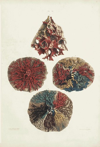
'Ducts and Glandules'
Regarding the figure on the right:
"Ducts injected more minutely with yellow, red, green, blue and black [wax]. This preparation shows two additional circumstances: -First, the glandules from which the ducts begin are seen filled with wax. Secondly, at the lower part of the preparation the separate ducts are seen passing above and beneath each other, to render the breast a cushion; whilst at the upper part the ducts are single."
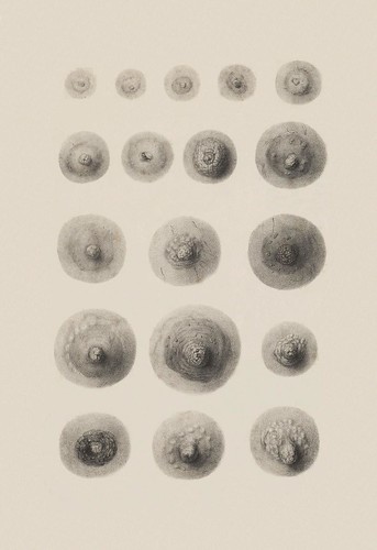
'The Nipple in its Changes'
These figures represent subjects aged from two through to old age. The examples in the first row are from pre-pubescent girls. Various influences - pregnancy, lactation, multiple or no offspring and menopause - are said to affect the appearance of the remaining figures. A few of these nipple drawings were derived from cadavers. It all seems fairly random, although obviously meant - from the accompanying notes - as a general guide to physiological and appearance changes.
[this plate in particular has been extensively background cleaned]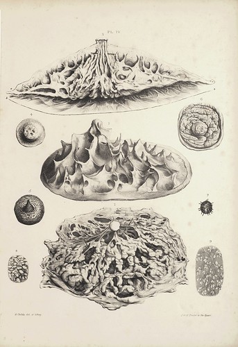
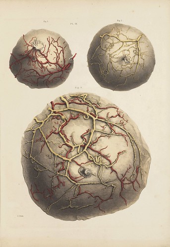
'Ligamenta Suspensoria and Sections'
Seen most specifically in the middle figure,
ligamenta suspensoria (aka Cooper's ligaments
*, after the author of this book) is a network of fibrous connective tissue throughout the breast. It provides structural support for
all the anatomical components and is responsible for giving the breast its characteristic shape.
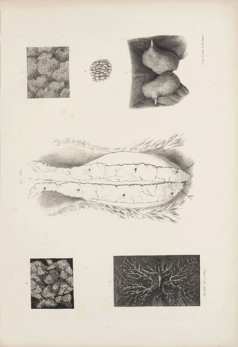
'Ducts, Glandules and Cells'
The top figure shows wax-injected milk ducts leading from small secretory glandules to the nipple. The bottom figure is a magnified version of one of those tracts and the middle drawings show the glandules at higher magnification again.
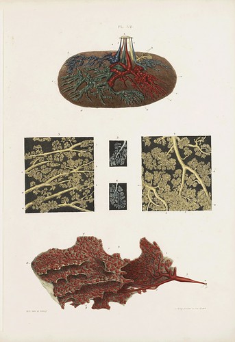
Arteries and Veins
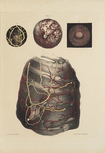
Arteries and Veins
In both of these images, arteries are shown in red and veins in yellow. That large figure at bottom in the second image is a complicated drawing of a subject facing towards us. It is attempting to show the route of both shallow and deep blood vessels in relation to the ribs. A section of the collar bone can be seen at the top and the edge of the sternum is on the right: therefore, this is a view of the circulation in the right breast. (the nipple is of course the white circle near the bottom of the image)
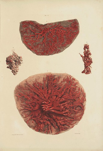
'Ducts, Reservoirs and Glandules'
The lactiferous tubes (milk ducts), injected with wax, in a deceased subject who was lactating at the time of her death.
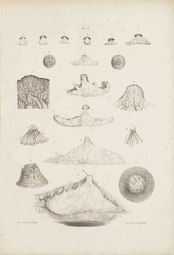
'Showing first the size of the gland at different ages'
The plate shows the cross sectional appearance of the mammary gland from about one to eleven years of age (across the top row). The central large figures on the page depict the gland size in subjects from age thirteen (at the top) to twenty four years. Details on either side show the magnified foliate appearance of the papillae of the nipple. There are also drawings of dissected ducts at the nipple terminus and sections showing the incredible abundance of minute blood vessels at the nipple surface.

"This plate is intended to show the external appearance of the nipple in the
male at different ages, the internal appearance of the gland as covered by its fascia at different periods of life, the glands and the ducts of the male gland injected, and the gland and ducts of the foetus"
The illustrations with dark backgrounds are ducts of the male mammary gland after they have been injected with mercury which helps, like the wax in other images, to delineate and visualise the micro-structure.
[all illustrations above are of human anatomy]
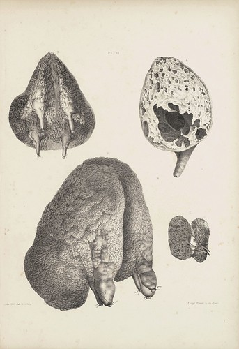
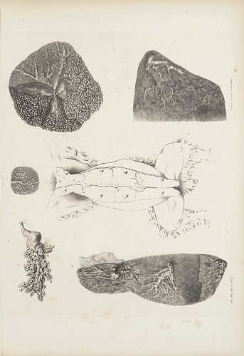
'The Mammary Gland of the Porpoise'
"Divided into two glands, one placed on the right, and the other upon the left side of the anus and vulva."
"Professor Owen informs me that some milk which he obtained from a porpoise felt like butter upon the tongue."
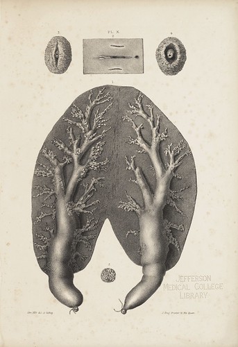
'The Dug of the Ass'
"Shows the two teats of this animal’s udder. The teats at first sight appear single; but one in this injected dug contained three mamillary tubes, and the other contained only two. The glands are injected with wax, and form a foliage upon their surfaces. Glandules appear upon every part of this foliage, and in these the milk-cells are readily traced. At the roots of the teats are reservoirs, of large size, but not proportionably equal in magnitude to those of the cow, yet still capable of containing many ounces of milk." {the drawings at the top are of a deer's udder}
[see:
Anatomy of a Mammary Gland in reference to animals]
'The Guinea Pig and the Cat'
The top three figures are from the guinea pig.

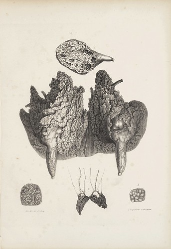
'Showing the Udder of the Goat and the Mammary Gland of the Rhino'
The top left image (magnified milk ducts and milk cells) and the bottom image (pair of teats, each with twelve mamillary orifices) belong to the rhino.
'Shewing the Udder of the Ewe'
"There are in the ewe two teats, leading into two large glands, and there are sometimes imperfect teats behind. [..] The milk tubes of the teats open into a reservoir, capable of containing many ounces of milk, and a mucous membrane lines it, similar to that which lines the teat. Milk canals begin from the reservoirs, and these form a foliage on the surface of the gland."
Of the Milk of the Ewe: "It is abundant, and is sometimes used as the food of children. It forms a considerable quantity of cream. Its butter retains a large quantity of curd, and therefore it easily becomes rancid. Its cheese is rich but contains much oily matter."
'Of the Mammary Gland in the Rabbit'
'On the Anatomy of the Breast' 1840, by Sir Astley Paston Cooper, is hosted by Jefferson Digital Commons from Thomas Jefferson University in Philadelphia. The 2-volume book can be downloaded either as two large pdf files or each section/illustration plate can be downloaded separately. All the plates (extracted from the pdfs) - which includes a few more than those seen above - have been uploaded to
this flickr set.
Sir Astley Paston Cooper
(1768-1841) had a distinguished academic and surgical career in London. He made significant contributions to the anatomy of cerebral circulation, vascular surgery, amputation and hernia repair techniques and was appointed Professor of Comparative Anatomy at the Royal College of Surgeons (where he also had two terms as President). He published a number of surgical/anatomy books and his renown as a surgeon was rewarded with a royal appointment and knighthood.
One of the characteristics that set Cooper's work apart from his peers was his rigid scientific approach, which included, in the case of his treatise on breast anatomy, only reporting findings related to specimens and subjects that he himself had examined and prepared. So perhaps my "random" comment up above is out of place: in the introduction to this work he advocates that all surgeons ought to follow such rigorous practices to give greater legitimacy to their published findings.
That he was able to obtain human specimens
at all in the early 19th century meant that Cooper had to deal with the so-called
"resurrection men" who robbed graves to supply medical schools. Back then, the only cadavers legally available for research included criminals sentenced to death and dissection by courts.
'On the Anatomy of the Breast' was the first detailed analysis of the anatomy and physiology of the breast and was Cooper's last book.
- Biography: one; two.
- Just because it hit the radar: "The Global Library of Women’s Medicine has launched online and is freely available in beta at www.glowm.com. More than 650 world experts have provided a definitive resource on the latest therapeutic options in women’s medicine for doctors as well as for women who are concerned about their health. Medical professionals who register have access to additional resources." [via]
- In general: links/summaries/bookmarks/feeds/twitter*new*


